Every day, modern laboratories produce thousands of microscopic images. You might view the cover glass as a simple, disposable consumable. In reality, it acts as a highly precise, final optical element in your microscopic imaging pathway. Misjudging your glass specifications leads to two incredibly costly outcomes. First, it causes severe optical aberrations in high-resolution fluorescence and confocal imaging. Second, it triggers workflow paralysis in automated digital pathology labs due to equipment jams and broken slides.
Procurement managers, lab directors, and lead researchers need a scalable, reliable strategy. Our goal is to provide a data-backed decision matrix. We will help you select the exact glass specifications required for your distinct instrumentation and diagnostic workflows. You will learn how to balance optical precision, automated handling, and long-term archival stability seamlessly.
Key Takeaways
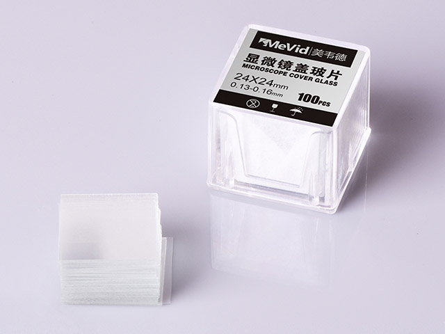
The 0.17mm Standard is a Composite: Standard No. 1.5 thickness (0.17mm) accounts for both the glass and the mounting medium between the glass and the specimen.
NA Sensitivity is Drastic: Objectives with a Numerical Aperture (NA) greater than 0.4 are exceptionally vulnerable to thickness variations; at NA 0.95, a mere 0.01mm error can degrade image intensity by 55%.
Scalability Requires Strict Tolerances: For high-throughput labs, prioritizing ISO 8255-1 compliant glass with HGB-1 hydrolytic resistance guarantees automated handling without sticking and ensures long-term slide archiving.
Application Dictates Shape: Beyond thickness, choosing between square, rectangular, and circular formats is strictly dictated by the imaging environment (e.g., automated slide scanners vs. live-cell culture wells).
The Optical Reality: Why Cover Glass Thickness Makes or Breaks Resolution
Microscope objective lenses are not magical tools. Manufacturers design them expecting a specific optical path length to achieve perfect focus. The glass actively corrects light pathways before they ever enter the objective lens. Using the wrong thickness fundamentally alters this path length. It introduces severe spherical aberration. This aberration causes light rays from different parts of the lens to focus at different points. The result is a hazy image and a massive loss of contrast.
We must deconstruct the common 0.17mm (No. 1.5) standard. Many laboratory technicians mistakenly believe 0.17mm refers exclusively to the physical glass itself. In truth, 0.17mm represents the total physical distance from the top of the cover slip down to the specimen. If you mount a biological specimen in a thick layer of aqueous liquid, you increase the overall path length. In these scenarios, you might need a thinner glass (like No. 1) to compensate for the liquid layer and achieve optimal focus.
Common Mistake: Relying blindly on No. 1.5 glass for every application without considering the depth of your mounting medium. Thick mounts demand thinner glass.
The established thresholds for thickness sensitivity are drastic. High numerical aperture (NA) lenses capture wider angles of light. This makes them incredibly sensitive to path length errors. We can observe the quantitative evidence in the chart below.
Objective Numerical Aperture (NA) | Thickness Deviation | Approximate Image Intensity Loss |
NA ≤ 0.4 (Low Magnification) | 0.01mm - 0.02mm | 0% (Largely Immune) |
NA 0.85 (High Magnification) | 0.01mm | 19% Loss |
NA 0.95 (Very High Magnification) | 0.01mm | 55% Loss |
As the table demonstrates, stringent thickness control becomes absolutely non-negotiable for high-end applications.
Step 1: Matching Glass Specs to Objective and Immersion Environments
Your choice of objective lens directly dictates your glass requirements. We must evaluate the distinct dynamics between dry lenses and immersion lenses.
Dry objectives observe specimens through air. Air has a refractive index of roughly 1.0. Glass sits at a refractive index of approximately 1.52. This harsh refractive mismatch makes dry objectives highly susceptible to thickness variations. Light bends aggressively at the air-glass interface. Any deviation in glass thickness amplifies this bending error, destroying your resolution.
Oil immersion lenses operate differently. They are much more forgiving if your mounting medium matches the borosilicate cover glass refractive index (~1.52). Immersion oil fills the air gap, creating a continuous optical path. However, a hidden danger exists. If you observe specimens in aqueous media (like saline) via oil lenses, the water creates a new refractive mismatch. Even under oil, thickness accuracy remains critically important for aqueous samples.
High-NA lenses frequently feature thickness correction collars. You can manually adjust internal lens elements to compensate for variations. Explain this operational workflow to your imaging staff. First, set the collar to 0.17mm and focus the microscope. Next, turn the collar slightly and refocus. Observe if the image contrast improves or degrades. Because real-world specimen preparations tend to run thick, adjusting the collar toward higher values (0.18–0.23mm) is often your optimal starting point.
Step 2: Selecting Microscope Cover Glass Types by Shape and Application
Shape dictates functionality in the laboratory. Exploring different microscope cover glass types allows you to connect specific geometries directly to laboratory applications.
Square: This format serves as the baseline for routine histology, cytology, and general non-automated microscopy. Dimensions like 22x22mm offer ample coverage for standard manual workflows.
Rectangular: These extended sizes (such as 24x50mm) are essential for whole-slide mounting. They easily cover large tissue sections and blood smears. More importantly, rectangular shapes ensure seamless compatibility with automated coverslipping machines.
Circular: You will find circular formats mandatory for precision positioning. They fit perfectly inside multi-well plates, confocal dishes, and live-cell imaging setups where standard rectangular slides cannot be used.
You must also weigh fixed tissue against live-cell considerations. Fixed tissue relies comfortably on standard No. 1.5 coverslips mounted on traditional slides. Live-cell imaging introduces distinct challenges. Cells must remain viable and stationary during prolonged observation. This typically requires specialized glass-bottom dishes. Researchers routinely coat these dishes with adhesion proteins, such as poly-D-lysine. These coatings promote cell attachment and maintain strict focal stability.
Best Practice: Always audit your vessel dimensions before ordering circular glass. A minor 1mm sizing error will prevent the glass from seating flat in a culture well.
Evaluating Scalability: Automation, AI Pathology, and Archiving
Procurement managers must look beyond basic optical clarity. Frame your purchase as a strategic investment in artificial intelligence and digital pathology readiness. Digital slide scanners utilize AI algorithms to stitch thousands of individual images together. These algorithms require completely uncompromised focal planes. Cheap, warped glass creates uneven topographies. This significantly increases scanning rejection rates and forces technicians to perform manual rescans.
High-throughput laboratories rely heavily on effortless automation. Autostainers and coverslipping machines use sensitive suction cups to lift and place glass. You must evaluate surface smoothness, strict dimensional cutting, and anti-sticking properties. Rough edges or sticky surfaces cause multiple sheets to lift simultaneously. This leads to broken slides, lost tissue samples, and costly equipment downtime.
Archival reliability represents another massive hurdle. Clinical laboratories must often store patient slides for decades. Enter the HGB-1 medical-grade hydrolytic resistance standard. Glass naturally reacts to moisture over time. Low-quality glass undergoes alkaline extraction, becoming cloudy or hazy. HGB-1 certified glass resists moisture degradation effortlessly. It ensures legal and clinical compliance in long-term slide archiving.
We strongly recommend building a strict compliance framework for vendor selection. Shortlist only those vendors that transparently provide ISO 8255-1 standard certifications. You can evaluate a supplier's commitment to these rigorous manufacturing standards by reviewing their cover glass quality control history.
Implementation Risks: Tolerances, Quality Control, and Handling
Academic and clinical labs frequently fall into the batch variability trap. Standard off-the-shelf optical cover slips feature surprisingly broad thickness variance from one box to the next. You might calibrate your system perfectly on Monday, only to experience severe spherical aberration on Tuesday after opening a new box.
For high-end confocal or super-resolution applications, standard ranges simply fail. We recommend upgrading to "High Tolerance" (1.5H) glass. Standard No. 1.5 glass fluctuates between 0.16mm and 0.19mm. The premium 1.5H designation tightens the manufacturing variance to a strict ± 0.005mm (0.165mm to 0.175mm). This upgrade eliminates focal drift during complex Z-stack imaging.
Elite facilities do not blindly trust new vendor batches. They actively verify tolerances using stringent Quality Assurance (QA) validation methods:
Precision Micrometers: Technicians use specialized jaw micrometers to perform multi-point thickness checks on random samples from every new shipment.
Interferometry: Advanced research centers utilize light wave interference technology. This non-destructive method offers extreme measurement precision for super-resolution demands.
Proper handling maintains optimal integrity. Implement these actionable handling best practices across your laboratory staff.
Store glass boxes in low-humidity environments. Desiccators prevent moisture buildup, which causes individual sheets to stick together.
Use lint-free cleaning methods. Standard paper towels leave microscopic debris that disrupts digital scanner auto-focus systems.
Never touch the center surfaces. Fingerprints deposit natural skin oils. These oils actively alter the local refractive index and introduce image artifacts.
Conclusion
Choosing the correct specifications directly impacts your diagnostic accuracy and operational throughput. You can streamline your procurement strategy by following a simple shortlisting logic. First, confirm your objective NA and your immersion type. This determines your exact thickness requirements. Second, select the shape based on your specific vessel or slide scanner geometry. Finally, filter your vendors by ISO compliance, HGB-1 hydrolytic resistance, and strict tolerance guarantees (e.g., 1.5H). This ensures your glass supports automated workflows seamlessly.
We advise buyers to take immediate action before committing to bulk contracts. Request sample batches and run them directly through your automated coverslippers. Perform internal micrometer checks on these sample lots. Verifying precision upfront protects your laboratory from downstream failures, ensuring perfect microscopic images every time.
FAQ
Q: What is the standard thickness of a microscope cover glass?
A: The industry standard is No. 1.5, which measures 0.17mm. Standard manufacturing tolerances usually range between 0.16mm and 0.19mm. For demanding high-resolution applications, labs utilize high-performance "1.5H" glass. This tightens the tolerance to a strict ± 0.005mm, ensuring perfect focal alignment.
Q: Why is borosilicate cover glass the industry standard?
A: It provides a specific refractive index of roughly 1.52, matching immersion oils and standard microscope objective lenses perfectly. Furthermore, it offers exceptional optical clarity and high chemical resistance against harsh laboratory solvents and mounting media used in slide preparation.
Q: How do you measure optical cover slips accurately?
A: Laboratories use precision jaw micrometers to take physical measurements across multiple points on the glass surface. For ultra-precise, non-destructive quality assurance, manufacturing facilities employ optical interferometry. This uses light waves to map out microscopic thickness variations flawlessly.
Q: Do I need No. 1 or No. 1.5 cover glass for aqueous samples?
A: It depends on your sample depth. While objectives are designed for 0.17mm (No. 1.5), this measurement includes both the glass and the liquid above the specimen. Using thinner No. 1 glass (0.13-0.16mm) often serves as a practical hack to compensate for thick water layers in fresh wet mounts.


